Onchocerciasis and Mycosis
Onchocerciasis
Introduction
Onchocerciasis, also known as "river blindness", is a disease caused by the parasite worm Onchocerca volvulus. It is transmitted to humans by the bite of infected black flies (Simulium spp.) that breed in rapids and streams, mainly in remote rural areas near fertile agricultural lands. This disease is mainly spread out in tropical areas where more than 99% of infected individuals are found in a large number of countries in sub-Saharan Africa, as well as several scattered sources in Latin America, including Brazil, Guatemala, Mexico and Venezuela.
In Venezuela, Dr. Potenza reported a case of onchocericiasis for the first time in 1949, diagnosing it in a nodule found in a patient from Monagas State. From then on, many cases of onchocerciasis began to be reported throughout the country. Epidemiological investigations were carried out, the results of which up to 1974 delineated two major sources along the Coastal Mountain Chain; one in the eastern part of the country (part of Monagas, Sucre and Anzoátegui States) and another in the Central States of Aragua, Miranda, Carabobo, Guarico, Yaracuy and Cojedes.
Dr. Convit's Studies
In 1960, Dr Convit took part in the fight to control onchocerciasis, a disease which by then had already become a public health problem because it is a common cause of blindness in the affected individual. For his work, he used a methodology similar to the control of leprosy to determine the epidemiological extent of the problem that covered nine states and the Amazon Territory of the country. Thirty thousand patients were diagnosed and given treatment and subjected to medical supervision.
Thus, Dr. Convit made important contributions in connection with onchocerciasis, carrying out research, supporting studies of this disease in the National Institute of Dermatology of the Vargas Hospital of which he was the Chief of the Dermatology Service, as well as the disclosure of data associated with the advances and updates on river blindness in national congresses, workshops and conferences.
Information for patients
How do you get onchocerciasis?
Onchocerciasis is caused by the parasite worm Onchocerca volvulus. This worm enters the body through the bite of tiny insects (1-5 mm) or simullidae (Simulium genus), commonly known as black flies. These flies develop their larval stage in rapid flowing rivers and clean streams. The female fly is infected when it bites an infected person to feed on blood, and then ingests microfilariae of the parasite (small worms). Once inside the fly, these microfilariae develop into infective larvae (third-stage larvae), which can be inoculated in a new healthy host when the infected fly seeks a blood meal again.
What are the signs and symptoms of onchocerciasis?
Once the worms enter the host, they provoke a reaction that causes them to be encapsulated in fibrous tissue or nodules on the skin, where they live and reproduce, generating the release of microfilariae which can move throughout the human body through the subcutaneous tissue. The symptoms of this disease are caused mainly by the presence of these microfilariae, which generate intense inflammatory responses, especially when they die. Infected people may have symptoms such as itching, rashes, swelling, and various skin lesions. In the majority of cases, there are nodules under the skin. Some infected people develop ocular lesions that can cause visual disability and permanent blindness.
How is onchocerciasis diagnosed?
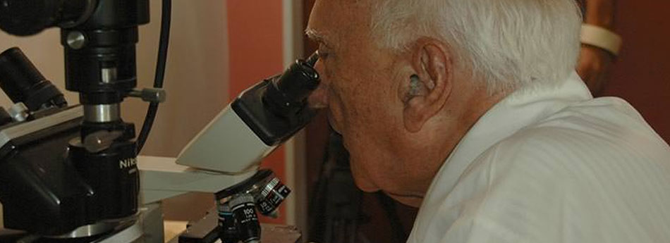
The simplest method of diagnosis is the direct method, which consists in collecting a small sample of skin and placing it in a drop of saline solution, and observe under the microscope how the microfiliarie abandon the skin in a few seconds, if it tests positive for the disease. The parasitological diagnosis confirms the clinical and epidemiological diagnosis by identifying the ocular and cutaneous lesions in individuals who live in or have recently visited the areas endemic for this disease. Microfiliarie can also be detected in blood by essays of PCR coupled to an ELISA or Southern blot to detect DNA (genetic parasite material), and an ELIS with recombinant antigens for the detection of anti-O volvulus antibodies in blood. These last methods of diagnosis are used widely and mainly to determine the epidemiological parameters, such as the prevalence and incidence of exposure to the parasite by the human population, as well as for monitoring programs of onchocerciasis control applied in endemic areas.
How is onchocerciasis treated?
Unfortunately, until now there is no vaccine or medication to prevent infection by O. volvulus. However, the WHO recommends the treatment of onchocerciasis with ivermectin at least once a year for about 10 to 15 years. An alternative to the ivermectin is diethylcarbamazine, taken following the same criteria. However, it is best that you undergo specialized medical consultation (dermatology) to verify the diagnosis and therefore receive the most appropriate treatment.
As part of the effective boost treatment for this disease, a nodulectomy (surgical removal of the nodules) is performed to remove the palpable subcutaneous nodules and eliminate the adult parasites of O. volvulus. This procedure, by eliminating adult parasites, diminishes the duration of the treatment. However, it is not always feasible due to the multiple lesions in each patient and its high cost.
What should I do if I think I have onchocerciasis?
The first thing you must do is go to your physician to be referred to a specialist dermatologist if necessary. In Caracas, you can go to the Dr. Jacinto Convit Institute of Biomedicine where there are specialists that handle consults and offer guidance to patients with onchocerciasis or who suspect they have it. If you live in the interior of the country you can get information at the nearest health center about the Dermatological Health service nearest to your locality to be evaluated by specialists.
Mycosis
Introduction
Mycoses, or fungal infections, are caused by different groups of microscopic fungi that are pathogenic for humans and multiply on the surface of the skin and in some organs. Depending on the tissue affected, mycoses can be categorized as superficial, subcutaneous, deep or systemic and opportunistic.
Superficial mycoses. In these fungal infections, the fungus infects only the superficial layers by invading the keratinized structures such as the stratum corneum, hair, nails and/or mucous membranes.
Subcutaneous mycoses. These affect the deeper layers of the skin associated with the dermis and epidermis.
Deep or systemic mycoses. These are the most serious infections caused by fungi as they affect the internal organs, where they reproduce.
Opportunistic mycoses. Also known as secondary systemic infections, they develop mainly in immunosuppressed persons or those who are undergoing prolonged antibiotic therapy.
The causative agents of fungal infections in general are everywhere; however, fungal infections are reported more frequently in tropical and subtropical countries with warm, humid climates, including Venezuela. They occur mainly in people who live in rural areas, where it is common to walk barefoot in their dwellings and therefore repeatedly suffer lesions from plants or other contaminated material. Additionally, these diseases can be contracted from infected animals such as dogs, cats, and other non-domestic animals. In general, these infections can affect persons of both sexes and all age groups without distinction. However, some types of fungal infections occur most frequently in adults, although children are exposed to the same environment.
In Venezuela, after the federal Sanitary Dermatological Services were established in 1970, they begin to register cases of fungal infections, both superficial and deep. Then in 1984 systematic studies of general fungal diseases began through the creation of working groups in several states of the nation under the name Grupos de Trabajo en Micología de Venezuela or GTMV (Mycology Working Groups of Venezuela. These groups provided a novel approach to the study of fungal infections, promoting advances in diagnosis, the discussion of the various forms of clinical presentation, the taxonomic study of fungi and experience in treatment, with special emphasis on endemic mycoses.
Dr. Convit's Studies
When the federal Services of Sanitary Dermatology began to register cases of mycosis in their clinics, Dr. Convit became involved in the studies of these diseases. Deeply committed to the study and cure of Hansen's disease, Dr. Convit developed work that identified the differences between this disease and some skin disorders, such as mycoses, which were often confused with the different types of leprosy. In addition, he participated in epidemiological studies to define the distribution of some mycoses in the country. Thus, his efforts contributed to the significant progress in this area.
Dr Convit made important contributions related to fungal infections by promoting courses on mycosis and dermatosis from the Dermatology Service of the Vargas Hospital and the Association for Dermatologic Research. He carried out research projects, evaluated new proposals for treatment as well as made public the data associated with the progress and updates in national congresses, workshops and conferences.
Information for Patients
In which parts of the body may mycoses appear?
Taking into account the different types of mycosis there are, these can be located basically in any part of the body. When they affect the scalp, they manifest themselves as dandruff or seborrheic dermatitis. On other occasions, when they affect the skin, blemishes appear with more or less pigmentation than the rest of the skin as well as different sizes. They can also cause cracks between the fingers and toes, round and reddish lesions anywhere, and inflammation in the armpits and groin areas. A fairly common type of mycosis is onychomycosis, which is difficult to eradicate, and affects toenails, and less frequently, fingernails.
What are the signs and symptoms of mycoses?
Corporal mycoses appear as visible, inflammatory lesions, often with specific characteristics associated with the fungus that produces them. In general we can say that fungal infections produce intense itching, blemishes and alterations in skin color, peeling of the skin, small blisters, whitish plaques attached to the mucous membranes of the mouth and genitals, and sometimes secretions associated with the local lesion.
How are fungal infections diagnosed?
In each of the types of mycosis the diagnosis is made primarily in clinical form by a doctor through the observation of the symptoms, which is usually sufficient to establish the diagnosis. However, it becomes necessary to identify the fungus that causes the disease so as to be able to provide the most appropriate treatment to the patient. The samples for analysis are generally secretions or tissues collected from the lesion, and in the case of systemic infections, x-rays are required. The evaluation of the sample is done mainly by direct microscopic study, although it's also feasible to carry out the isolation of the causative agent in special culture media and/or detection of specific antibodies by double immunodiffusion, the latter mainly in cases of systemic mycoses. Histopathological analysis is used sometimes to confirm the diagnosis, although it is not a routine procedure.
How are fungal infections treated?
The most advisable treatment for superficial mycoses is the application of topical antifungal creams, ointments, lotions, powders and sprays. Currently, the topical product most used due to its high degree of effectiveness is clotrimazole, a broad-spectrum antifungal derived from imidazole. If this treatment is not effective, the dermatologist must be consulted for a more thorough assessment and prescription of oral medication. Attention to injuries, coupled with good hygiene habits allows faster healing and prevents re-infection. It is important to always remember that before self-medicating yourself, you should consult your physician. In terms of deep mycoses, it is essential that they be treated as soon as possible by the dermatologist specialist, who can recommend local, topical or oral antifungal treatment according to each case, and in some cases will indicate surgical procedures to remove lesions or drain abscesses.
What should I do if I suspect I have some sort of fungal infection?
The first thing you should do is go to your doctor to be referred to a specialist dermatologist if required. In Caracas, you can go to the Dr. Jacinto Convit Biomedicine Institute, where there are specialists who handle consults and offer guidance to patients with various skin conditions. If you live in the interior of the country, you can find information at your local health center on the Dermatological Health service nearest to your locality to be evaluated by specialists.
